Astaxanthin–Powerhouse Antioxidant

Astaxanthin–Powerhouse Antioxidant

Astaxanthin-The Powerhouse Antioxidant

19 Known Medical Benefits of Astaxanthin

Astaxanthin Shown to Slow Brain Aging

I have recommended Astaxanthin for many years before it gained its notoriety as it has now. Natural astaxanthin is the most powerful antioxidant known to science, and has the ability to optimize the health and radiance of the skin by providing protection and support to all layers.

Astaxanthin is the most powerful of over 600 different types of Carotenoids which are pigments in plants, algae, and photosynthetic bacteria. These pigments produce the bright yellow, red, and orange colors in plants, vegetables, and fruits; and are responsible for the “pinkish” color of both shrimp and salmon

I initially recommended it for brain health in my effort to ward off Alzheimer Dementia years before the symptoms may appear. Now it is recommended for a host of other health issues including reducing the cytokine storm that had once resulted from COVID,

There are over 114,000 articles about Astaxanthin on PubMed–the go to site comprising of more than 32 million citations for biomedical literature life science journals, and online books.

I do not use Mercola’s brand of Astaxanthin but have nothing against it. I use a brand from Allergy Research mostly because of the source of the ingredients(AstaZine), the delivery system and the cost of the product itself. Either way, Astaxanthin should be on your list of what I call the “dailies”—supplements you should consider taking every day

I am currently doing a study on Ascorbate Acids role in balancing the neurotransmitters in the brain which I will pass over to you once it is done and I am satisfied with the results. This will be a part of a new website titled Strategic Supplementation which God willing,  I  plan to launch in 2021.

Good ol’ Glutathione – the Antidote to Snake Oil “Detoxes”

Good ol’ Glutathione – the Antidote to Snake Oil “Detoxes”

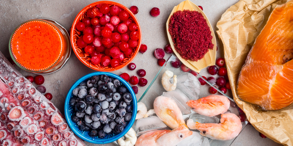
When patients make the decision to clean up their diet and put more effort into establishing and maintaining healthy lifestyle habits, many of them find benefit in starting out with some type of guided cleanse or detox. Television and magazine ads provide no shortage of slick programs promising miraculous transformations in health and physique through following these strategies, which typically range anywhere from a week to a month in duration. No doubt, clever marketing and pretty packaging can convince people who are fatigued, overweight, and living with chronic pain, that the answer to their woes lies in subsisting on cabbage soup, green smoothies, or a diet made up entirely of raw foods.

Patients would come in with drops, tablets, pills all claiming miracle detoxification.  The glitz and glamour these quick-fixes are wrapped up in obscure, the simple and perhaps even boring truth: the body “detoxes” itself constantly. Compounds that are produced by the body, itself, albeit with the aid of accessory nutrients, are the most effective things for internal housekeeping, and these cleansing processes take place all the time, with no need for drastic measures like drinking copious amounts of lemon juice, maple syrup and cayenne pepper. Through eating a whole-foods, nutrient-dense diet, patients can support their body’s primary antioxidant and detoxification powerhouse: glutathione

Glutathione does not appear out of nowhere. It is manufactured by the body and is considered the mother of all antioxidants. Until recently, the only way to supplement our own reserve of glutathione was through IV injections. The enzymes responsible for its synthesis and recycling require several vitamin and mineral cofactors. The short list includes magnesium, riboflavin and selenium. Magnesium is needed for the synthesis of glutathione, itself. Selenium is required for glutathione peroxidase (GPx), which converts potentially harmful hydrogen peroxide into water, leaving behind oxidized glutathione (GSSG). At least five variants of GPx are known to be selenoproteins: GPx1 (in the cytosol), GPx2 (specific to epithelial cells in the lungs and intestinal lining), GPx3 (thyroid and kidneys), GPx4 (phospholipid-hydroperoxide), and GPx6 (active in the olfactory epithelium). Moving along in the glutathione redox cycle, riboflavin, as part of flavin adenine dinucleotide (FAD), is used by glutathione reductase, which restores oxidized glutathione into reduced glutathione (GSH).

  • GSH resides in the cell  —  There are approximately 50-72 trillion cells in the body
  • GSH is the mother of all antioxidants
  • It is considered the strongest antioxidant in the world because it is what your own body produces
  • Glutathione is a tri-peptide meaning it has 3 amino acids

The name, glutathione, is indicative of the presence of at least two of its components: the amino acid glutamine, and the presence of sulfur (thio), in the form of the amino acid cysteine. Glycine is the third amino acid that makes up the glutathione tripeptide. Being that it is a tripeptide, it’s especially amusing that many of the popular quick-fix cleanse and detox programs marketed to uninformed consumers are low in protein. While plant foods do, of course, provide protein, vegetable-based proteins are typically not as bioavailable nor as complete as animal proteins. Programs that call for the elimination of animal foods, at least, temporarily, run the risk of not providing the body with enough of the amino acids it needs to support the synthesis of its own in-house detoxifying substances, of which glutathione is only one.

  • It is 5000 times stronger than Vitamins C & E
  • Vitamin C has 5 extra electrons to donate
  • Vitamin E has 3 extra electrons to donate
  • GSH has 1 million extra electrons to donate

Although glutathione is synthesized by the body, there are health situations that may benefit from supplementation. Specifically, these include conditions associated with rampant oxidative stress, in which the use of glutathione might outpace production, or which may be caused or exacerbated by reduced levels of this key antioxidant. For example, cytosolic glutathione levels are markedly reduced in the substantia nigra of patients with Parkinson’s disease. Damage to mitochondria is believed to underlie several other neurodegenerative conditions, such as Alzheimer’s disease. Increasing glutathione levels in the brain is being explored as a therapeutic adjunct for slowing or preventing Alzheimer’s and its precursor, mild cognitive impairment.

Supplemental glutathione is sometimes an appropriate choice for patients, but can be difficult to deliver to the body. Until recently, the only way to supplement our own reserve of glutathione was through IV injections. As a peptide, glutathione administered orally would be broken down during digestion. Providing precursor molecules, such as N-acetyl-cysteine, or glutathione in its reduced form or as S-acetyl-glutathione, in which the acetyl group protects the compound from being degraded in the GI tract, are ways to boost levels of this crucial compound.

  • Raising cellular glutathione increase the release of toxins
  • Toxins are most always attached to fat molecules such as bile
  • GSH forces toxins to attach to fats (bile) which is then returned to the Liver
  • How your body rids itself of toxins is directly related to NOT getting Cancer
  • You can never downgrade inflammation without cellular detoxification

*If you check Pub-Med, as of 922/15 there are 321,156 studies done on Glutathione for most every disease complex

*Also on PubMed as of 9/22/15 there are 6,217 research articles on Glutathione’s affect on aging

Here is an excellent link from Huffington Poast on Glutathione–Mother of all Antioxidants

In my result driven practice, I use Glutathione supplements which absolutely, undoubtedly work as they should. Please contact me at [email protected] or call 619-231-1778

Vitamin D-Comprehensive

It is only within the last few years that the important role Vitamin D plays has been researched and recognized.

Here are some facts regarding “Vitamin D” which is actually a hormone as opposed to a vitamin.
* Vitamin D, also called Calciferol, is a fat-soluble vitamin which means it is found primarily in animal fat. Unlike other vitamins that must be obtained solely through supplements or diet, vitamin D is manufactured in the body when sunlight comes in contact with the skin.

Here is a short list of what Vitamin D is responsible for.

  • Vitamin D is responsible for regulating over 800+ genes in the body!
  • It is a known fact that Vitamin D 25- Hydroxy levels lower than 15  will lead to many varieties of Cancer especially Colon Cancer
  • Vitamin D deficiency was responsible for older patients losing their balance and falling.  Two studies, one from the Journal of the American Geriatrics Society and the other in Pharmaco Economics support this theory
  • .Low Vitamin D associated with age related cognitive decline, dementia and Alzheimers.    In a new study published earlier this month in JAMA Neurology, researchers demonstrated a significant association between vitamin D insufficiency and cognitive decline that is specifically seen in Alzheimer’s disease and dementia. The results reinforce the importance of identifying vitamin D insufficiency among the elderly. Here, low vitamin D levels were associated with significantly faster rates of decline in memory and executive function performance..
  • Vitamin D has historically been used to treat Low Blood Pressure as there appears to be a correlation between blood pressure and low blood calcium levels and Vitamin D is directly responsible for where calcium should be and should not be.
  • Vitamin D is vital for bone growth and repair, especially in children and the elderly. Calcium and phosphorous levels in the blood stream are maintained by vitamin D. Vitamin D assists in the absorption of these important nutrients during digestion. This role in calcium and phosphorous absorption makes vitamin D an important part of bone growth and maintenance; and liver and kidney health Vitamin D also contributes to the function on the thyroid gland and to the immune system.

*The vast majority of the vitamin D used in the human body is manufactured in the skin. When vitamin D is consumed it is absorbed into the blood stream from the intestines with the help of bile. From the intestines Vitamin D is transported to the liver where it may be stored or used by the body. The daily Recommended Dietary Allowance (RDA) of vitamin D is 400 IU’s for an average adult man/woman.

Why suddenly is everyone Vitamin D deficient?

  • Not eating the foods high in Vitamin D which include fish liver oil, egg yolks, butter and cod liver oil. Vitamin D which naturally exists in milk is destroyed during the pasteurization process making it necessary to replace the naturally occurring Vitamin D that once was in the milk with synthetic Vitamin D which the body cannot utilize
  • Not enough sun exposure due to fear that the sun causes cancer when in fact that is not the case. Poor diet is!
  • Actually,  it may not be a deficiency of Vitamin D atoll, but a deficiency of Vitamin F, Vitamin D’s antagonist. Vitamin D picks up calcium from the gut and puts it into the blood. Vitamin F takes it from the blood and puts it into the tissues.

How to Optimize Vitamin D?

  • To optimize your levels, you need to expose large portions of your skin, such as your back, chest, legs, and arms, to sensible sun exposure. And, contrary to popular belief, the best time to be in the sun for vitamin D production is actually as near to solar noon as possible.
  • During this time you need the shortest exposure time to produce vitamin D because UVB rays are most intense at this time. Plus, when the sun goes down toward the horizon, the UVB is filtered out much more than the dangerous UVA.
  • Just be cautious about the length of your exposure. You only need enough exposure to have your skin turn the lightest shade of pink. Once you reach this point your body will not make any additional vitamin D due to its self-regulating mechanism. Any additional exposure will only cause harm and damage to your skin.

Vitamin D is important, but as we know, it is not the only key player. It is also important to assess calcium, magnesium, vitamin K2, and other trace minerals for deficiencies and support accordingly. There are several labs that can assess RBC minerals and fat-soluble vitamins.

What about Vitamin F–I never heard of this vitamin before?

  • Vitamin F is a source of essential polyunsaturated fatty acids which are needed to transport calcium from the blood to the tissues.
  • It contains Linolenic and Linoleic Acids which the liver will convert to Arachidonic Acid.  Arachidonic acid is the only form of polyunsaturated fat that can be utilized by the body.
  • It does not exist in vegetable oil—only in fat meat and butter—not margarine.
  • Because polyunsaturated fats reduce blood-cholesterol levels, they are much preferred over saturated fats.

D without F will cause the blood calcium level to increase at the expense of the tissue calcium level because Vitamin D not only picks up calcium from the gut in a deficiency of F, it brings calcium from the tissues back into the bloodstream and then targets the additional D to be stored by the Liver.

Vitamin F Deficiency results in or plays a major role in the following conditions::
* Hives                                     *Dry Skin
* Itchy Skin                              *Muscle Cramps (Charley Horse)
* Canker Sores (Herpes)        *Sun Poisoning
* Hypothyroidism                     *Sun Sensitivity
* Ridged Nails                          *Heat Prostration
* Poor Hair Quality                   *Prostate Problems

In my practice, I often recommend a blood test called Vitamin D 25-Hydroxy to determine the blood levels of Vitamin D.. Once the blood Vitamin D levels are determined, I may recommend specific supplements;hopefully on a short rather than long term basis.  I do not want my patients to stay on supplements forever, and prefer they increase their D levels using foods and the sun.

Note: Because vitamin D is stored in the body (in the liver), it can be toxic if taken in excess quantities. Vitamin D doses of three of four times the RDA may result in nausea, excessive thirst, confusion, heart rhythm abnormalities, weakness and headaches. Vitamin D is also known to interfere with certain medications including steroids and some seizure drugs. Point being do not take Vitamin D because it is a new fad–see a health care provider that knows what they’re doing, get the lab test and correct appropriately

There is a lot of chatter going on regarding Vitamin D.  For further information on the subject, feel free to contact me at [email protected] or 619-231-1778

Poor Sleep = Alzheimer’s

Poor sleep associated with buildup of toxic Alzheimer’s protein

Restful sleep is required for us to store and save our memories. If you are not getting enough sleep each night, you may be at an increased risk of developing Alzheimer’s disease. This is something that I feel equally applies to many other chronic diseases as well.

I previously wrote about the connection between Sugar and Alzheimers with Alzheimers often referred to as Type 3 Diabetes.  I am worried about this for myself and my patients. This article is a continuation of additional information and insights to help us never to get Alzheimers /Dementia to begin with.

I have also mentioned using sleeping medications does not allow the brain to go into the correct and proper REM cycle and therefore you may in fact be sleeping but your brain is not regenerating and rebuilding as intended.

In a recent study published in Nature Neuroscience, researchers at University of California, Berkeley, found evidence that poor sleep, specifically a deficit of deep sleep, is associated with a buildup of the beta-amyloid protein. Excessive deposits of beta-amyloid are the primary suspects in the pathology of Alzheimer’s disease, as this toxic protein ends up attacking the brain’s long-term memory.

This correlation between sleep, beta-amyloid, memory, and Alzheimer’s disease has been growing stronger. Sleep is when our body repairs itself. Quality sleep prevents these toxic proteins from accumulating and destroying brain cells. A buildup of beta-amyloid protein has been found in Alzheimer’s patients as well as in patients with sleep disorders. A study from University of Rochester in 2013 found that the brain cells of mice shrunk during non-rapid-eye-movement (non-REM) sleep to free up space for the cerebrospinal fluid to wash out toxic metabolites such as beta-amyloid protein.

Overall, the results of the new study demonstrated that the more beta-amyloid you have in certain parts of your brain, the worse your memory. In addition, the less deep sleep you get, the less effective you are at clearing out beta-amyloid protein. Researchers do not know yet which of these two factors – the poor sleep or the build-up of beta-amyloid protein – begins the cycle that triggers this cascade.

This is a new pathway linking Alzheimer’s disease and memory loss, which is significant since we can do something about it, since poor sleep is treatable and can be improved by modifying sleep habits. It is important that you go to sleep around the same time every night. When the timing of your sleep is shifted even if the duration of sleep is the same, it’s not going to be as restorative. In addition, avoid watching TV or using your computer before bed. Computer screens (smartphones and laptops) emit light in the blue part of the spectrum. This doesn’t cause a problem during the daytime, but at night, this blue light limits the production of melatonin. As a result, it disturbs your sleep-wake cycle. There are free apps you can install on your computer if you absolutely need to be on your computer at night that adjusts colors in a way that reduces the stimulating effects of blue light at night.

Caffeine and other stimulants can also keep you up and interfere with sleep. It is best to avoid these four to six hours before bedtime. Finally, try to workout earlier in the day. Exercise increases cortisol and can make falling asleep very difficult.

If behavior and lifestyle modifications are not enough, there are nutrients and botanical agents that can significantly promote restful sleep. Melatonin is a hormone whose primary role is in controlling the body’s circadian rhythm. While adequate levels of melatonin are essential for quality sleep, its production declines significantly as we get older, often causing sleep difficulties associated with aging. Thus, supplementing with melatonin has been shown improve sleep quality. In addition, 5-HTP can further support endogenous melatonin production during the night to help with staying asleep.  Inositol is a member of the B vitamin family that promotes relaxation and helps maintain the proper metabolism of serotonin. In addition, L-theanine provides calming neurotransmitter production clinically proven to reduce stress and improve the quality of sleep.

Valerian root, German chamomile, passion flower, and lemon balm are all calming botanicals used for centuries to help with insomnia. They have all been shown to decrease the amount of time it takes to fall asleep as well as improve sleep quality.

However, although taking herbs to aid in sleep is far better than toxic pharmaceuticals, the bottom line is to find out why we are not sleeping well to begin with.

In addition to eliminating sugar and foods that break down into sugar, I highly recommend Meditation twice daily, Qigong and the powerful Mindfulness Technique.  Doing the above is almost a sure shot of getting a good nights sleep.  So take one of these up, practice makes perfect and before a year is out, if not sooner, you will be sleeping beautifully and as such re-setting our brains circuitry.

B12-

B12 – the Vitamin for All Ages

We have long appreciated the role of particular vitamins in contributing to the physical structure of the body. There’s vitamin D for healthy bone turnover, and vitamin C for hydroxylation of proline and lysine, required for the synthesis of collagen. Vitamin B12 is another of these key factors, and it contributes to the building and maintenance of myelin, the protective covering that surrounds neurons. Severe B12 deficiency results in a number of signs and symptoms that affect multiple body systems. Among these are the hematological changes categorized as pernicious and megaloblastic anemias, decreased methylation and elevated homocysteine, and compromised neurological function. The last of these is especially concerning, because some of the adverse effects may be irreversible, even upon repletion of B12 stores.

One group commonly at risk for B12 insufficiency is the elderly. Many older people may be taking stomach acid blocking medication, which will inhibit the body’s ability to absorb this vitamin. Additionally, due to decades of guidance from government nutrition authorities to reduce consumption of foods rich in saturated fat and cholesterol, people may be avoiding some of the primary dietary sources of B12, including red meat (liver, in particular), egg yolks, and shellfish. (Older people might also steer clear of these foods if they find them more difficult to chew, and reduced stomach acid will make them harder to digest, as well.)

It is especially troubling that older people may be deficient in this nutrient, because B12 is critical for healthy cognitive function. It is well known that a B12 deficiency can result in memory loss, disorientation, and dementia. It would be tragic if some older patients were misdiagnosed as having Alzheimer’s disease when the cause of changes in their cognition and behavior was actually a vitamin deficiency. Researchers say that low vitamin B12 status “is an important risk factor for loss of brain volume in older community-dwelling adults. These findings suggest that plasma vitamin B12 status may be an early marker of brain atrophy and thus a potentially important modifiable risk factor for cognitive decline in the elderly.” (Emphasis added.)

Compared to patients with other types of dementia, Alzheimer’s disease patients have lower levels of B12 in their cerebrospinal fluid. The resulting reduced availability of methyl groups is believed to lead to decreased synthesis of myelin, membrane phospholipids, and the neurotransmitter  —all of which would have disastrous consequences for cognitive function. After recognizing that elevated plasma homocysteine was associated with cognitive decline in older adults, researchers who set out to determine whether it was insufficient folate or insufficient B12 that was responsible, found that it is B12 that influences healthy cognition. A doubling of holotranscobalamin (bioavailable or “active” B12) concentration was associated with a 30% slower rate of cognitive decline, whereas a doubling in homocysteine or methyl-malonic acid (markers for B12 insufficiency) was associated with greater than 50% more rapid cognitive decline.

Other groups that may be at risk for low B12 status are vegetarians and vegans, since the most concentrated sources of this nutrient are animal proteins. Lacto-ovo vegetarians may get sufficient amounts from eggs and dairy products, but those who avoid animal-sourced foods altogether may have difficulty remaining nutrient replete without careful supplementation. Children of strictly vegetarian parents have experienced developmental difficulties resulting from decreased myelination due to B12 deficiency. These include severe psychomotor retardation, frontoparietal cranial atrophy, and the more general catch-all, “failure to thrive.” Most disturbing is that even when hematological signs of deficiency are corrected, there may be some degree of long-lasting neurocognitive defects later in child development. Researchers familiar with B12 deficiency in infants emphasize that strict vegetarian parents should be made aware that some of the resulting compromises in development during fetal and neonatal life may be irreversible, and, thus, it would be wise to take precautions to ensure adequate maternal B12 levels during pregnancy and breastfeeding.

 NOTE: Many of you have heard me talk about “Aunt Sadie” who beginning in the 1950’s gave B12 injections to our entire family and everyone else who lived in the neighborhood.  She had a booming B12 practice.

Alzheimer’s Disease: Type 3 Diabetes?

Alzheimer’s Disease: Type 3 Diabetes?

It is no coincidence that we are witnessing a skyrocketing increase in the incidence of Alzheimer’s disease (AD), which parallels those of metabolic syndrome, type 2 diabetes, and obesity. All of these are, in part, outcomes related to carbohydrate intolerance and the mismatch between our biological makeup and our modern diet and lifestyle. In fact, the connections between glucose, insulin dysregulation and Alzheimer’s disease are so strong that many researchers now commonly refer to AD as “type 3 diabetes

The blood-brain-barrier is a powerful border that carefully regulates the entry of fuel substrates and nutrients from the periphery. However, it is not capable of protecting the brain from the deleterious effects of an onslaught of refined carbohydrates, oxidized vegetable oils, and nutritionally empty processed foods. The brain is an intensely energy-hungry organ, and anything that impedes its use of glucose—such as peripheral and/or central insulin resistance—will have disastrous consequences for cognitive function. Alzheimer’s disease is the end stage manifestation after a significant number of neurons have “starved to death” due to a loss of their ability to metabolize glucose.

Although the outward manifestations of AD—such as memory loss, confusion, and disturbing behavioral changes—are easy to observe, there are also physiological factors that can be measured and quantified. One of the earliest and most profound observable biochemical changes in the AD brain is a reduction in the rate at which the brain uses glucose, called the cerebral metabolic rate of glucose (CMRglu). This can be measured in vivo, with AD patients showing upwards of 45% reduction in CMRglu compared to healthy, age-matched controls. Some researchers see this decline in glucose usage by the brain as the predominant abnormality in AD

Interestingly, the decline in CMRglu can be observed in people at risk for AD (based on family history or genotype) as early as in their 30s or 40s, long before overt signs of AD have manifested. Thus, the decreased CMRglu can be seen as a kind of “canary in the coal mine”—an early warning sign that something is going awry in the brain. The extent of the reduction in CMRglu is tied to AD severity. A longitudinal study using PET scan to measure CMRglu.  in people age 50-80 showed that reduced hippocampal CMRglu at baseline predicted progression from normal cognitive function to AD, with the greatest reductions at baseline correlating with the quickest development of full-blown AD.

At baseline, hippocampal glucose metabolism in people who progressed from healthy to AD was 26% below that of people who did not develop AD, and the annual rate of decline averaged 4.4%. In people who progressed from normal to mild cognitive impairment (a precursor to AD), CMRglu was 15% reduced at baseline, with an annual rate of decline at 2.4%. The rate of decline for people who had normal CMRglu at baseline and did not develop AD was just 0.8%. Assuming the rates of decline were somewhat constant, extrapolating backward indicates that the decline may have started as early as 20 years before overt signs of AD were present. At baseline, despite the already decreased CMRglu in some subjects, all subjects were cognitively normal. This suggests that a starting point of reduced glucose usage in the brain and a stronger rate of continued decline might be one of the first triggering events in AD. The brain may be able to compensate for years before damage is so widespread that overt symptoms are observable. The normal forgetfulness and foibles we associate with “just getting older”—Where did I leave my keys? Don’t I have an appointment somewhere this week?—might be the earliest indicators that the brain is struggling to fuel itself.

An interesting potential contributor to the reduced CMRglu is peripheral and/or central insulin resistance. Plasma concentration of insulin is positively correlated with AD severity.  When neurons become insulin resistant, they are afflicted by the same pathology that occurs in the periphery—an inability to properly metabolize glucose, causing glucose to accumulate in extracellular spaces for an extended period of time. This results in rampant glycation and the formation of advanced glycation end products (AGEs). These AGEs add insult to injury by forming cross-linkages with each other that may alter the shape of neuronal synapses and impede cellular communication and nerve impulse transmission in the brain, with cognitive abnormalities being an obvious consequence. With hyperinsulinemia affecting 40% of people over age 80, it’s no surprise to find a link between insulin dysregulation and a condition that preferentially strikes older individuals. Moreover, hyperinsulinemia has been found to be and independent risk factor for AD.

The beta-amyloid (Aβ) plaques often implicated as a cause of AD may, in fact, be a result of peripheral hyperinsulinemia. In addition to the reduced CMRglu, the presence of insoluble Aβ plaques is one of the defining signatures of AD pathology. However, Aβ is a normal product of protein degradation, and there is no evidence that AD patients overproduce Aβ. Rather, the problem seems to be that Aβ isn’t cleared away as it should be, which results in these small, otherwise soluble peptide fragments aggregating into insoluble plaques. (These plaques are then subject to glycation and blocking synapses, adding yet another obstruction to neuronal communication.)

A fascinating thing to note is that what is responsible for clearing away Aβ in a timely manner—before it dwells long enough to form plaques—is insulin degrading enzyme (IDE), the same enzyme that clears away insulin. However, the affinity of IDE for insulin is so high that even small amounts of insulin completely the degradation of AB. One study demonstrated that peripheral infusion of insulin in older subjects increased the level of AB in cerebrospinal fluid within 120 minutes, and this also correlated to decreased memory function. Thus, the formation of Aβ plaques is facilitated by hyperinsulinemia. Adding yet another piece of evidence to the theory that Aβ plaques are an effect of AD pathology, rather than its cause, is the fact that the decline in CMRglu precedes the formation of the plaques. Therefore, the presence of Aβ plaques is not likely the triggering factor. (They may exacerbate disease severity, but they are not the initial event in its initiation.)

Considering the connections between impaired glucose metabolism, chronically elevated insulin, and Alzheimer’s disease, the phrase “type 3 diabetes” is viable.

5-HTP

Synthesized from the amino acid tryptophan, 5-hydroxytryptophan (5-HTP) is the rate-limiting precursor to serotonin and melatonin, our relaxation hormones that are important for proper sleep. 5-HTP supplementation has been shown to be useful in enhancing serotonin levels in humans, which is why it is most known for its role in helping with depression.

5-HTP has shown promise with sleep disorders and insomnia, especially increasing rapid eye movement (REM) sleep, thus improving slow wave sleep (SWS). In fact, some studies have also shown promise with improvement of childhood sleep terrors. Much of this makes sense, given 5-HTP’s involvement in the synthesis of melatonin, known to be one of the regulatory hormones involved in the sleep-wake cycle. Serotonin (5-HT), too, has been known for its powerful sedative effects, especially after ingesting the tryptophan in a huge Thanksgiving turkey meal (although this is most likely in large part due to overeating). According to an animal study, “The similarity of the effects of 5-HTP and tryptophan suggests that they both act as serotonin precursors.”

5-HTP is not found directly in foods, but made from individual amino acids. In order to be effective, 5-HTP must cross the intestinal lining and enter the bloodstream. In a healthy gut environment, absorption occurs easily. 5-HTP readily crosses the blood-brain-barrier, moving into targeted tissues where it is then converted into the active neurotransmitter, serotonin. Studies have found that, when taken with vitamin B6, 5-HTP facilitates the manufacture of serotonin, which increases melatonin production.

Fibromyalgia, insomnia and sleep terrors are the most common conditions studied in regards to 5-HTP supplementation and sleep. During a 90-day open trial, nearly 50 percent of the patients affected by fibromyalgia patients experienced significant improvement in quality of sleep, fatigue, anxiety and pain when taking 5-HTP. Other conditions in which fatigue is a primary concern may also benefit from a similar treatment.

Insomnia in children is quickly becoming a troubling issue, affecting nearly 20 to 30 percent of young children, and leaving a plethora of trailing health and behavioral consequences. Many young children with insomnia continue to have sleep issues later in life. Lack of adequate sleep in children is often linked to physical and learning disabilities, difficult temperaments, autism, epilepsy, and attention problems, among other things. Night terrors are a common cause for sleep issues in children between the ages of 3 to 12 years. These are episodes that usually cause screaming, but can also cause sudden awakening with persistent fear, sweating, confusion and increased heart rate. In one study, children with night terrors were given 5-HTP and compared to a similar group of children who were not given 5-HTP. The results of this study indicated 83.9 percent of the children treated with 5-HTP were episode-free after six months, thus showing a hopeful solution to this particular cause of sleep insufficiency in children. Another small case study tested the effect of 5-HTP on sleep in two children with schizophrenia. After treatment with 5-HTP, increased REM and sleep improvement was noted.

In consideration of the high impact sleep deprivation has on quality of life, behavior, mood and health, it is important that practitioners deal with foundational sleep issues when considering all health complications. 5-HTP provides a good starting place for addressing sleep issues in both children and adults and has a history of safe usage. Being a derivative of serotonin, not only will 5-HTP improve sleep quality, but it will also indirectly influence mood and behavior in a positive direction. This offers a lot of hope to patients and practitioners since mood and emotions impact the healing process and the perception of wellness. Many health conditions are rooted in sleep deprivation and 5-HTP offers a safe, easy starting point for health and wellness.

NOTE#! : In my practice I often recommend a 1mg Melatonin sub-lingual tablet (Source Naturals) not at bedtime but depending on the patient, 1-4 hours prior to bedtime but never at bedtime itself.  So if you go to bed at 10, I have them take the 1 mg Melatonin at 7, 8 & 9

I additionally recommend a 50mg 5-HTP to be taken 3 hours prior to bedtime

NOTE #2: Remember the reason why people can go to sleep initially, wake up and then cannot go back to sleep is because their blood sugar is plummeting during the night. As a result we are awoken from a sound sleep which immediately increase the Cortisol levels which in turn stabilize the blood sugar.  The down side to this is that if Cortisol increases Melatonin decreases and you cannot fall back to sleep 1,2,3.

NOTE #3: You are not awakening to pee, you are awakening because of blood sugar.  By the end of 2015 or early 2016, Alzheimers will officially be referred to as Type 3 Diabetes

NOTE #4: Another new phenomenon is children who have insomnia and whose parents are either giving them pharmaceutical medications for sleep or melatonin–both of which are abhorrently disgraceful

Tags: Sleep, melatonin, depression

Sleep Study Questionnaire

 Sleep Study Questionnaire

The purpose of this brief questionnaire is to accrue information now that may prevent us getting Alzheimer’s/Dementia in the future.

Alzheimer’s is now being referred as Type 3 Diabetes!!

Through the use of MRI’s andf other state of the art diagnostics, the above problems actually begin to show up in our 30s.

All of the questions below are approximates—please answer as such

  1. What time do you go to sleep at night  ____________
  2. Do you wake up during the night  ___________
  3. If so how many times do you wake up during the night  ______________
  4. Do you take Ambien, Xanax, Diazepam’s, Melatonin etc. to help provoke sleep  ______________
  5. If yes to the above, do you use every night or just some nights _____________
  6. Have you ever been officially diagnosed having sleep apnea  _______________
  7. Would you say that overall and in general, you get a good night’s sleep  ______________
  8. On a level of 1-10 how would you honestly rate your stress levels  ___________

Detox Tips – Long Term n Easy

Acetyl-Glutathione & Intracellular Detoxification

By Jack Tips (Ph.D, CCN)

Clinical nutritionists have long recognized the premier importance and vast significance of the molecule, glutathione (GSH), in human health.  It is the body’s most important antioxidant/detoxifier, and protects the cells from free radical damage incurred during mitochondrial production of adenosine triphosphate (ATP) energy. Other cellular free radical protectors are the enzymes catalase and super-oxide dismustase. Simply put, if glutathione levels drop, the body is prone to cancer, wasting disease, autism, hepatitis, cataracts, Alzheimer’s, Parkinson’s COPD, asthma, schizophrenia, bipolar and aging—just to cite a few conditions bearing numerous medical references

Raising glutathione levels in the cells has been a challenge.  Glutathione can be put into a supplement, but it’s difficult to get it through the intestines into the bloodstream.  Once in the bloodstream, it’s difficult to get supplemental GSH into the cells as degradation occurs in the bloodstream.  Studies showed taking supplemental GSH does not raise blood levels.  Thus oral supplements were deemed  to assist in the gastrointestinal tract; perhaps some help in the blood, but not enter the cells where it’s needed.  Intravenous GSH administration became the most effective way to push GSH into the cells.

Recently, a new form of GSH became available—one that demonstrates it can be absorbed through the G.I. tract and survive degradation in the bloodstream, cross cell membranes and thus favorably impact the cells, as well as help prevent cancer.  It is acetyl-glutathione featured in Systemic Formulas’ GCEL.

S-Acetyl Glutathione is an alternative frm of the reduced GSH. Several studies have shown that this precursor of GSH is well absorbed and more stable throughout the digestive tract than the reduced       L-glutathione and has a positive effect on many oxidative stress bio-markers.  S-Acetyl  Glutathione enters the cell directly and once inside the cell it is converted to GSH by the cytoplasmic thioesterases that remove the acetyl group thus rapidly raising Intracellular GSH levels.

Because of GSH ability to promote detoxification from within the cells, its supplementation can flood the detox pathways and cause large amounts to enter the small intestines via gall bladder excretion via the bile.  To prevent resorption from the intestines, practitioners utilize BIND (Toxin Elimination), a superactived charcoal, to absorb and bind toxins so they cannot be resorbed.  Together GCEL and BIND assist the body in “removing the cause” of inflammation, hormone resistance, cell membrane damage, and errant cell metabolic processes.

NOTE: In clinical practice, detoxification proponents often employ the 60 Day Systemic Detoxification Program to help the body clear out the interfering toxin backlog, improve liver function, open detoxification pathways, and impact cellular metabolism with protective antioxidants.  Then boosting GSH to drive the detoxification effort into the cellular processes offers the body an amazing opportunity to rejuvenate its cellular functions from which a more optimal health can be restored.  Systemic’s Intracellular Detoxification program provides state of the art true cellular detoxification support that serves the brain, cells, and innate healing processes.